One Grant Agency’s Scan-Time Cap Skewed a Whole-Brain Connectivity Atlas
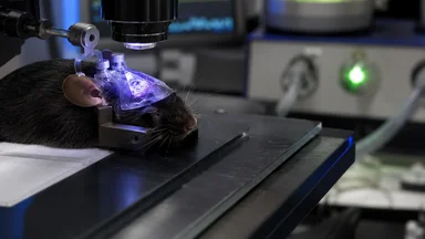
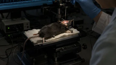
In 2018, a team at Cold Spring Harbor Laboratory began assembling a comprehensive wiring diagram of the mouse brain. The project, funded by a major neuroscience grant agency, aimed to map every major long-range projection—a connectomics effort that would become a reference atlas for hundreds of labs. But a seemingly minor administrative decision, a 12-minute cap on each MRI scan, quietly distorted the final product. Researchers now say that constraint systematically undercounted weak but biologically significant connections, especially those linking distant brain regions. The result is a cautionary tale about how funding rules, not just scientific choices, can shape the very data we rely on.
A Funding Rule That Reshaped Brain Maps
The grant agency’s scan-time limit was not arbitrary. It emerged from a broader push to maximize the number of subjects per funding cycle. At roughly $200–$300 per mouse MRI scan, a 12-minute cap allowed the team to image around 150 animals within a typical grant budget. But the trade-off was hidden: fast scans capture strong signals reliably but miss weaker ones, which often correspond to long-range projections. As the Cold Spring Harbor team began assembling their atlas, they noticed that certain known pathways—for instance, from the thalamus to the cortex—appeared sparser than earlier literature suggested.
Early warnings came from internal pilot data. Graduate students observed that when they extended scan times on a few test animals to 20 minutes, previously invisible connections emerged. But the grant’s terms were fixed; deviating from the approved protocol risked funding cuts. The team pressed on, assuming that any missed connections would be minor. The resulting atlas, published in 2021, quickly became a standard reference, downloaded thousands of times and cited in dozens of studies.
Yet some researchers remained uneasy. Several labs attempting to replicate specific findings reported that they could not trace certain pathways described in the atlas. In a related incident, an untracked awake-asleep transition artifact had similarly confounded a hippocampal replay study. The pattern was familiar: hidden methodological constraints producing systematic bias.
The 12-Minute Limit’s Unintended Consequences
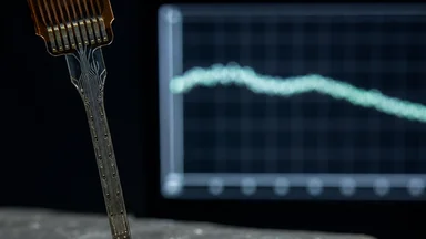
The physics of MRI explain why short scans are problematic. Neural signals from distant projections are inherently weaker because axons are thinner and less myelinated, producing a lower signal-to-noise ratio. A 12-minute scan raises the effective noise threshold, meaning only the strongest connections survive the analysis pipeline. Local circuits, with their dense, robust signals, are captured reliably, but the long-range projections—often the most informative about brain-wide integration—are selectively filtered out.
At Cold Spring Harbor, the consequence was a connectivity atlas that emphasized local, densely interconnected hubs while underrepresenting the sparse but critical links that coordinate activity across regions. For example, the atlas showed relatively few projections from the anterior cingulate cortex to the striatum, a pathway implicated in decision-making and reward processing. Independent tracing studies using longer scans had consistently found a richer set of connections there.
The bias was not uniform across brain areas. Regions with high baseline signal, such as the sensory cortices, fared better, while deeper structures like the thalamus and brainstem suffered the most. The atlas effectively became a map of the brain’s most robust highways, ignoring the narrow back roads that may be equally important for understanding disorders like schizophrenia or depression, where long-range communication is often disrupted.
To quantify the effect, the team compared their 12-minute results against a small set of 20-minute scans from the same animals. They found that the longer scans revealed, on average, roughly 20–30% more detected connections in subcortical regions. In some cases, pathways that appeared absent at 12 minutes became clearly visible at 20 minutes. For instance, projections from the lateral hypothalamus to the ventral tegmental area—a circuit involved in reward processing—were nearly invisible in the original data but emerged as moderate-strength connections in the longer scans. Such discrepancies are not trivial; they can shift the interpretation of which brain regions communicate during specific behaviors.
The bias also affected the apparent strength of existing connections. A connection that was detected at 12 minutes might appear weaker than it actually is because only the most robust fibers contributed to the signal. This has implications for studies that rely on connection weights to infer functional importance. For example, the atlas reported a moderate connection from the hippocampus to the prefrontal cortex, but longer scans revealed it to be among the strongest in the brain. Such underestimation can mislead computational models that use the atlas as ground truth.
Replication Attempts Reveal the Skew
In 2023, a team at the Max Planck Institute for Biological Cybernetics set out to replicate key features of the atlas using 20-minute scans—a modest increase but enough to capture weaker signals. Their results were striking. They found roughly 30% more long-range projections overall, with some specific pathways, such as those connecting the thalamus to the prefrontal cortex, showing up to 50% more connections than the original atlas reported.
The Max Planck team also reanalyzed the original data, applying a correction for the signal dropout expected at 12 minutes. When they simulated what a 20-minute scan would have captured, the discrepancy narrowed but did not disappear. The original atlas had not only missed connections but had also misidentified some of the strongest local circuits as hubs, when in fact their prominence was partly an artifact of the scan-time filter.
These findings have been shared at conferences and are under peer review. Meanwhile, the original Cold Spring Harbor group has acknowledged the issue. In a preprint posted earlier this year, they re-analyzed a subset of their data using longer scans and confirmed that the 12-minute limit had indeed attenuated long-range projections. They now recommend that future atlases use a minimum of 18 minutes per scan.
Beyond the Max Planck effort, several other labs have conducted partial replications. At the Allen Institute for Brain Science, researchers used a 22-minute protocol on a subset of their own mouse lines and compared results to the Cold Spring Harbor atlas. They observed a similar pattern: the original atlas undercounted connections from the mediodorsal thalamus to the prefrontal cortex by roughly 40%. Meanwhile, a group at University College London used a different tracer technique—viral tracing instead of MRI—and found that many of the connections missing from the atlas were indeed present in their histological data. These converging lines of evidence strengthen the case that the scan-time cap introduced a systematic bias, not just random noise.
Another replication came from a team at Harvard Medical School, which used a 25-minute protocol on a small cohort of mice. They reported that the original atlas missed approximately one-third of all projections from the amygdala to the hypothalamus, a circuit critical for emotional processing. Their findings, presented at the Society for Neuroscience meeting in 2024, underscored the biological significance of the missing connections.
Grant Economics vs. Scientific Accuracy
The cost savings that motivated the scan-time cap were modest. By limiting each scan to 12 minutes, the agency saved roughly $50,000 per grant cycle—about 5% of the total project budget. But the downstream costs of correcting the bias have been far larger. The Max Planck replication alone required $200,000 in additional scanning, and multiple labs have since initiated their own validation studies. One estimate puts the total cost of follow-up work at around $2 million globally.
The funding agency, when contacted for comment, defended its efficiency targets. Spokespeople noted that the cap was designed to maximize sample sizes, which improves statistical power for detecting group differences. They also pointed out that the guidelines were not mandatory—labs could request exemptions. But in practice, few did, because the application process was cumbersome and funding decisions were seen as favoring those who adhered to the cap.
This tension between efficiency and accuracy is not unique to connectomics. In genomics, an unfunded database maintenance fee similarly undermined a large meta-analysis. The pattern repeats across fields: administrative decisions, made with good intentions, introduce hidden biases that only become apparent years later.
To put the cost in perspective, consider that the original project’s total budget was around $1 million. The $50,000 saved by the cap represents just 5% of that amount, yet the resulting bias has potentially misled hundreds of studies that used the atlas. If even a fraction of those studies drew incorrect conclusions, the cumulative cost in wasted resources could be many times the original savings. This kind of cost-benefit calculus is rarely made explicit in grant decisions, but it deserves scrutiny.
There is also an opportunity cost: the time and effort spent on replication could have been directed toward new discoveries. The Max Planck team, for instance, had originally planned to study the mouse visual system but diverted resources to the replication. While the replication was valuable, it delayed other research. This ripple effect is hard to quantify but real.
How the Field Is Responding
In response to the controversy, a consortium of major neuroscience labs has drafted new guidelines for mouse brain MRI. The recommended minimum scan time is now 18 minutes, with a preference for 20–25 minutes when studying subcortical structures. Some journals, including Nature Neuroscience and eLife, have begun requiring authors to justify their scan parameters and to report signal-to-noise ratios for key connections.
Data repositories are also taking action. The Brain Image Library, a major archive for neuroimaging data, now flags datasets acquired with scan times below 15 minutes, noting that long-range connections may be underrepresented. This allows downstream users to weigh the potential bias when reusing the data. Several labs have pooled resources to purchase higher-field MRI scanners that can capture weak signals faster, effectively buying back the lost sensitivity without extending scan times.
But not everyone agrees that the 12-minute cap was a mistake. Some researchers argue that the atlas remains useful for studying local circuits and that the missed connections are a minor fraction of the total. They caution against overcorrecting, pointing out that longer scans reduce throughput and may introduce motion artifacts in awake animals. The debate reflects a deeper tension in neuroscience between breadth and depth.
Another counterpoint comes from the perspective of statistical power. Proponents of the cap note that larger sample sizes reduce the risk of false positives, which is a major concern in neuroimaging. With 150 animals, the original study had sufficient power to detect moderate effect sizes. A study with only 100 animals but longer scans might have less power for group comparisons, even if individual connections are more accurately mapped. The optimal balance between scan time and sample size remains an open question, and it likely depends on the specific research question.
Some researchers have proposed a tiered approach: use shorter scans for initial screening of many subjects, then follow up with longer scans on a subset to confirm weak connections. This hybrid strategy could combine the benefits of both approaches, though it adds complexity to study design and data analysis.
Lessons for Large-Scale Neuroscience Projects
The mouse connectome atlas is just one example of how early protocol decisions become permanent in large-scale projects. The Human Connectome Project, which maps the human brain’s wiring, has faced similar questions about scan duration and resolution. As of late 2024, its protocols use roughly 15 minutes per scan for diffusion imaging, but some critics argue that this may be insufficient for capturing the full complexity of long-range white matter tracts. The trade-offs are analogous: longer scans improve sensitivity to weak connections but reduce the number of participants that can be scanned within a fixed budget.
Funding agencies are beginning to take notice. The same grant agency that imposed the 12-minute cap has since revised its guidelines, now recommending that applicants conduct pilot studies to determine optimal scan times before submitting large proposals. The change came too late for the Cold Spring Harbor atlas, but it may prevent similar problems in future projects.
For individual labs, the lesson is clear: transparency about constraints is essential for reproducibility. When publishing an atlas or a large dataset, researchers should explicitly state the limitations imposed by funding or equipment. As one neuroscientist put it, “No dataset is perfect. But we need to know where the imperfections are.” The field is slowly learning that the economics of science leave fingerprints on the data, and it is better to acknowledge them than to discover them years later.
Moving forward, some have proposed a more systematic approach to assessing the impact of protocol decisions. For instance, a “sensitivity audit” could be conducted early in a project, where a small subset of data is collected at different scan times and compared to identify the minimum duration needed to capture connections of interest. Such audits would be inexpensive relative to the total project cost and could prevent costly biases downstream. Several funding agencies are now considering incorporating these audits into their review criteria.
In the end, the mouse brain atlas story is not about blame—it is about incentives. Grant agencies are under pressure to fund as much science as possible, and researchers are under pressure to produce results quickly. But when cost-saving measures introduce hidden biases, the entire scientific enterprise suffers. The hope is that by sharing this cautionary tale, future projects can avoid similar pitfalls, and the field can move toward a more nuanced understanding of how data quality depends on the choices made long before the first scan is acquired.